Posts

Image
Day 19: oxygen glucose deprivation experiment on C17.2 cells - I labelledup the plates with either Control, 1.5 hr OGD or 3 hr OGD. - Obtain the 1.5hr plate and the 3hr plate and aspirate the media from them. Wash each well out with 500µl of PBS and aspirate that out as well to ensure that there is no more media left in the wells. - place 500µl of the artificial CSF in both the 1.5 and 3 hour plates. - I took both plates and place them into separate hypoxia chambers. Start with the 1.5hr plate. Flood the chamber with nitrogen for one minute and clasp the tubes shut when complete. - Place the chamber back into the incubator and set the timer for 90 minutes. - Obtain the 3 hour plate and place it into the other hypoxia chamber. Also flood this chamber with nitrogen for one minute, clasp the clips shut and place it into the incubator. Set the timer for 3 hours. - while the oxygen glucose deprivation is happening, prepare the peptides as follows: Dilution of the pept...

Image
Day 18: LDH, Hoechst and MTT assays to assess the cytotoxicity of TAT, Myr-p110 and TAT-P110 from 0µM – 10µM Aim: to assess the cytotoxicity of varying concentrations of the peptides TAT, Myr-p110 and TAT-p110 by assaying LDH, Hoechst and MTT with DMSO Reason for assaying LDH: LDH is lactate dehydrogenase which catalyses the conversion of pyruvate -> lactate during anaerobic respiration. Anaerobic respiration only involves the process of glycolysis which occurs only in the cytosol. Thus, if the peptides became cytotoxic to the cells, then the lactate dehydrogenase would leak out into the medium and therefore be detectable by increased absorbance and less transmission of UV. Reason for assaying Hoechst: Hoechst specifically stains nuclei blue. If there are more nuclei present this means that less cells have died as a result of cytotoxicity. If there are less nuclei present this means that more cells have died as a result of cytotoxicity. It is required that we make a ...
Image
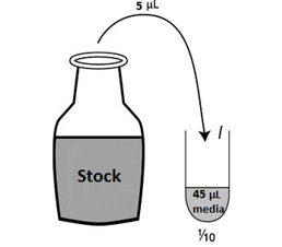
Day 17: Treating C17 cells with p110 peptide                        It was required that we diluted the stock solutions which were available to us to make up the desired concentrations for the wells: 0.1μM, 0.3μM, 1μM, 3μM, 10μM.   The TAT and Myr-p110 were both 10mM stock solutions whilst TAT-p110 was 4mM stock solution as it would not dissolve properly previously. The desired final concentrations meant that we had to dilute the stock solution into 1/10 and 1/100 solutions: -           Put 45 μl media into each of the tubes – the aim is to have 50 μl total volume by the end of the dilution. -           Take 5μl TAT Stock 10mM and place it into the 1/10 TAT tube. Vortex to ensure that the stock has dissolved properly. -         ...

Week 4: Inhibiting mitochondrial fission using p110 peptide varients in an attempt to prolong cell survival during neonatal hypoxic ischaemia

Image
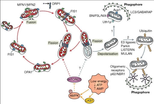
The theory behind the experiment: - There is an increase in mitochondrial mitophagy during neonatal hypoxic ischaemia. The cell becomes stressed as a result of the reduced oxygen in the environment. Reactive oxygen species (ROS) being formed after electrons react with molecular oxygen in the electron transport chain. The mitochondria which have become damaged will undergo the process of mitochondrial fission, mediated by the proteins Drp1, MFF, MID49/51 and Fis1. The mitochondria which have accumulated damage will have their damaged features taken to just one area in the mitochondrion. Once fission has occurred and one mitochondrion has become two, the mitochondrion which had accumulated its damage will undergo mitochondrial mitophagy. - As a result, less ATP is available for the cell but more ROS is produced. In addition, the mitochondrial membrane also becomes leaky releasing apoptosis stimulating factors and cytochrome c, therefore stimulating apoptosis and exaggerating th...
Day 15: Simply splitting cells I split cells from the two flasks that I left incubating last week and combined them into two flasks but with different media. One flask contained the media that I had previously used before doing my own oxygen glucose deprivation experiments while the other contained a different medium. On Monday (day 16) I will compare the confluence of the cells in both flasks and hopefully will be conducting more cell culture experiments next week!
Image
Day 14: Visualising Parkin and LC3B on western blot from p9 mice samples at 0 hours after 70 minute hypoxic ischaemia Result for Parkin: Parkin was visible at 52 kDa on the contralateral and ipsilateral lanes. The bands were more intense on the contralateral lanes which indicates that after 0 hours, there is not much recruitment of Parkin to the mitochondria for subsequent mitophagy. Result for LC3B: LC3B bands seemed to be at approximately equal levels in each lanes, but the mitochondrial lanes (last two lanes) are slightly brighter and this is most likely because the LC3B is starting to get recruited to auto phagosome and undergoing mitophagy.
Image
Day 13: Visualising PINK1 and LC3B, and running a western blot on 0 hour hypoxic ischaemic p9 mice samples The results for PINK1 and LC3B: PINK1 and LC3B seemed to be observed more in the 24 hour lanes (last 4 lanes) than in the 4 hours lanes. This is understandable, as cells which underwent oxygen glucose deprivation for a longer period of time would be under more stress and consequently more mitochondrial mitophagy would occur in those cells. Running a western blot on p9 mice cell samples at 0 hours after hypoxic ischaemia for 70 minutes: I had previously tested for mitochondrial mitophagy quality control proteins on p9 mice samples at 2 hours and 24 hours, thus we needed to test for changes in the proteins at 0 hours. We decided to test for the presence of Parkin and LC3B. I followed the same western blot procedure that I had conducted before and left the membrane at 4 degrees in the cold room with the primary antibodies for visualisation after...