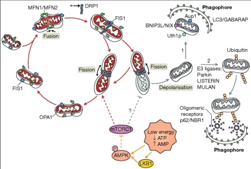
Day 14: Visualising Parkin and LC3B on western blot from p9 mice samples at 0 hours after 70 minute hypoxic ischaemia
 Parkin was visible at 52 kDa on the contralateral and ipsilateral lanes. The bands were more intense on the contralateral lanes which indicates that after 0 hours, there is not much recruitment of Parkin to the mitochondria for subsequent mitophagy.
Result for Parkin:

Result for LC3B:
LC3B bands seemed to be at approximately equal levels in each lanes, but the mitochondrial lanes (last two lanes) are slightly brighter and this is most likely because the LC3B is starting to get recruited to auto phagosome and undergoing mitophagy.


Comments
Post a Comment