Day 27 and 28: Assays, secondary antibody incubation and splitting cells: The assays: I ran assays for all of the OGD plates for Hoechst, MTT and LDH and the results were as follows: Secondary antibody incubation for immunofluorescence: I had to incubate the coverslips with secondary antibodies against the TOMM20 (which would be an indicator of mitochondrial morphology and presence) and Drp1 (which would be an indicator of fission. TOMM 20 were going to appear red whilst Drp1 was going to appear green on the confocal microscope. I incubated the coverslips in darkness for the rest of the day. At the end of the day, I mounted the coverslips onto slides which contained DAPI which would mark the nucleus and this would appear blue on the confocal microscope. Mounting the coverslips onto the slides: The slides were labelled with their relevant characteristic and the cloverslips were placed on top. The coverslips were secured in place with nailvarnish to the micro...
Popular posts from this blog
Week 4: Inhibiting mitochondrial fission using p110 peptide varients in an attempt to prolong cell survival during neonatal hypoxic ischaemia
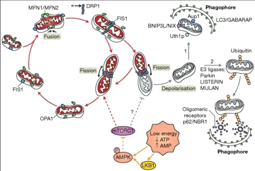
The theory behind the experiment: - There is an increase in mitochondrial mitophagy during neonatal hypoxic ischaemia. The cell becomes stressed as a result of the reduced oxygen in the environment. Reactive oxygen species (ROS) being formed after electrons react with molecular oxygen in the electron transport chain. The mitochondria which have become damaged will undergo the process of mitochondrial fission, mediated by the proteins Drp1, MFF, MID49/51 and Fis1. The mitochondria which have accumulated damage will have their damaged features taken to just one area in the mitochondrion. Once fission has occurred and one mitochondrion has become two, the mitochondrion which had accumulated its damage will undergo mitochondrial mitophagy. - As a result, less ATP is available for the cell but more ROS is produced. In addition, the mitochondrial membrane also becomes leaky releasing apoptosis stimulating factors and cytochrome c, therefore stimulating apoptosis and exaggerating th...
Week 3
Day 11: Splitting cells, my first mistake and calculating protein concentrations I managed to split the C17 cells on my own and unassisted from two previously split flasks. The aim was to keep the spare cells for oxygen glucose deprivation experiments and transfections with mCherry. What is mCherry? mCherry is a special flurophore which is used in molecular biology techinques as a tracer to track the localisation of molecules or organelles. In this case, we wanted to use it to view mitochondria. Unlike Keima or JC1 which I previously had experience with, mCherry would simply flurorece bright red once it was localised to mitochondria in the cells. The structure of mCherry: The structure of mCherry is such that it is a beta barrel motif with a fluorophore which is attached on the gap of the beta barrel. In addition there is also an alpha helix which runs down the beta barrel stabilising the chromophore in the centre. The theory behind using Cherry with re...


Comments
Post a Comment