Day 9: Oxygen/glucose deprivation to C. 17 cells
We wanted to observe the effect of oxygen glucose deprivation on the C 17 cells. The way in which we did this was by immersing the cells in artificial cerebrospinal fluid. The artificial cerebrospinal fluid did not contain any glucose and treated it with a vaccum to ensure that there was no oxygen in the fluid. we incubated two plates for 1 hour and a half and another two for 3 hours by placing it in a hypoxia chamber and flooding the chamber with nitrogen.
The result on the control plates:
The control cells did not under go oxygen glucose deprivation. The top photo shows the neurones with no marker dyes whilst the bottom shows that they were treated with markers JC1 and Hoechst. Hoechst stains the nuclei and chromatin. JC1 is a special fluorescent dye that changes colour depending on membrane potential. When the mitochondrial membrane potential is high, JC1 is aggregated and fluoresces red. However when the mitochondrial membrane potential is low, the JC1 forms monomers and fluoresces green.
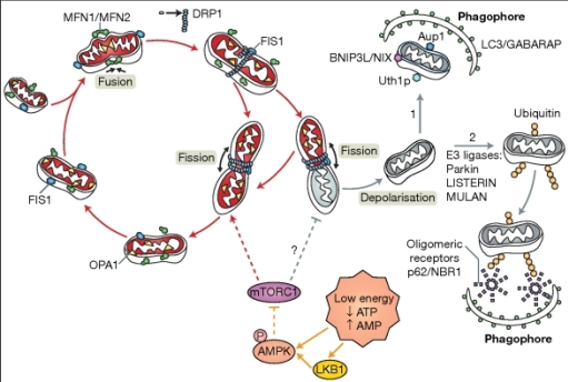
In healthy mitochondria, the membrane potential is high and when mitochondria are stressed the mitochondrial membrane potential decreases, indicating that mitophagy may follow. The control cells shows that there was a substantial amount of red fluorescence indicating that the mitochondria were not stressed and that JC1 was still in its aggregate form.
Upon oxygen/glucose deprivation:

:
There is significantly less red fluorescence in the plates exposed to oxygen glucose deprivation, indicating that the overall mitochondrial membrane potential was reduced.
We wanted to observe the effect of oxygen glucose deprivation on the C 17 cells. The way in which we did this was by immersing the cells in artificial cerebrospinal fluid. The artificial cerebrospinal fluid did not contain any glucose and treated it with a vaccum to ensure that there was no oxygen in the fluid. we incubated two plates for 1 hour and a half and another two for 3 hours by placing it in a hypoxia chamber and flooding the chamber with nitrogen.
The result on the control plates:
The control cells did not under go oxygen glucose deprivation. The top photo shows the neurones with no marker dyes whilst the bottom shows that they were treated with markers JC1 and Hoechst. Hoechst stains the nuclei and chromatin. JC1 is a special fluorescent dye that changes colour depending on membrane potential. When the mitochondrial membrane potential is high, JC1 is aggregated and fluoresces red. However when the mitochondrial membrane potential is low, the JC1 forms monomers and fluoresces green.
In healthy mitochondria, the membrane potential is high and when mitochondria are stressed the mitochondrial membrane potential decreases, indicating that mitophagy may follow. The control cells shows that there was a substantial amount of red fluorescence indicating that the mitochondria were not stressed and that JC1 was still in its aggregate form.
Upon oxygen/glucose deprivation:

:
There is significantly less red fluorescence in the plates exposed to oxygen glucose deprivation, indicating that the overall mitochondrial membrane potential was reduced.


Comments
Post a Comment