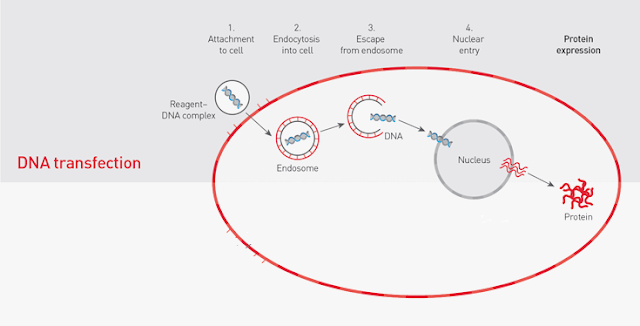
Day 8: Transfecting the C.17 cells
We decided to transfect the C.17 cells with lipofectamine 2000, which is a lipid rich mixture containing lipid micelles that a capable of transporting genetic information extra cellularly to the intracellular environment. The lipid micelles contain hydrophilic heads and hydrophobic tails that can fuse with the C. 17 cells.
What is Keima?

Keima is a special coral derived acid stable fluorescent protein which can fluoresce different colours depending on the pH that it is exposed to. The Keima cDNA is ligated into a plasmid with a mitochondrial tag at its N terminus. The DNA that we wanted to transfect was a plasmid called Keima.
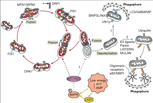
In order to understand how Keima works, it is important to get an insight on how mitochondrial mitophagy works
The mechanism of mitophagy (Exner et. al., 2012)
We decided to transfect the C.17 cells with lipofectamine 2000, which is a lipid rich mixture containing lipid micelles that a capable of transporting genetic information extra cellularly to the intracellular environment. The lipid micelles contain hydrophilic heads and hydrophobic tails that can fuse with the C. 17 cells.

The liposome has a cationic exterior and is able to attract the overall partial negative charge on the cDNA of the plasmid. Once inside, the liposome releases the plasmid which is taken up into the nucleus, where it is transcribed into its corresponding mRNA transcript. After post translational modification, the mRNA transcript is translated to the protein product: Keima.
What is Keima?
Keima is a special coral derived acid stable fluorescent protein which can fluoresce different colours depending on the pH that it is exposed to. The Keima cDNA is ligated into a plasmid with a mitochondrial tag at its N terminus. The DNA that we wanted to transfect was a plasmid called Keima.
In order to understand how Keima works, it is important to get an insight on how mitochondrial mitophagy works
The mechanism of mitophagy (Exner et. al., 2012)
 |
A) In normally functioning mitochondria, the mitochondrial membrane potential is high and PINK1 is continuously turned over by proteolysis and the levels of PINK1 on the mitochondrial membranes is low. (B) Low mitochondrial membrane potential indicates mitochondrial damage and PINK1 proteolysis is inhibited, causing its accumulation on the mitochondrial membrane. PINK1 recruits Parkin to the mitochondrial membrane and Parkin ubiquitinates the mitochondrial membrane proteins. Ubiquitin associates with the ubiquitin-adaptor protein, p62, and the phagophore engulfs the mitochondria, and upon the entry of lysosomes, the mitochondria are degraded.
Keima utilises the change in pH when the mitochondria is engulfed by the lysosome, to change its fluorescence. At the cell's physiological pH, ie. pH 7, Keima fluoresces green when it is localised to the mitochondria. When the autolysosome has engulfed the mitochondria during mitophagy, there is a pH change from pH 7 to pH 4. The drop in pH also causes Keima's fluorescence to change from green to red, thus being an effective indicator of mitophagy.

In order to see this in action, made up 6 control plates with 3 different volumes of DNA/Keima and 6 plates with an electron transport chain uncoupler, CCCP. It was expected that the plates treated with the CCCP would have more red fluorescence as a result of more mitochondrial mitophagy.
|

Comments
Post a Comment