Day 5: Identifying LC3II and Optineurin


What is
LC3II?
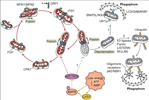
LC3II may also be referred to as LC3B is a 17kDa protein with
a corresponding isoform, LC3I (LC3A). During mitophagy, the autophagosomes are
recruited to the mitochondria which are destined to be degraded. The
autophagosomes engulf the mitochondria and at the same time, the LC3I which was
in the cytosol becomes conjugated to phosphatidylethanolamine to form LC3II
which is recruited to the autophagosomal membrane.
Lysosomes fuse autophagophagosomes to form autolysosomes –
the mitochondria is degraded by the lysosomal hydrolases and LC3II is degraded.
Therefore, by identifying LC3II using fluorescent markers (secondary
antibodies), we can detect the extent of mitophagy in the ipsilateral and
contralateral brain samples. It is the lysosomal turnover of LC3B that can
reflect the process of mitophagy as a result of hypoxic ischaemia.

Result for
LC3II localisation:

LC3II
was successfully located at 17kDa and ubiquitous protein GAPDH at 37kDa.
What is Optineurin?

After parkin has ubiquitinated outer membrane proteins on the
damaged mitochondria which is destined for auto lysosomal degradation, Optineurin
stabilises itself through its ubiquitin binding domain. When parkin is not
present, the Optineurin is only partially stable as it transiently associated
with the damaged mitochondria. Once the Optineurin has become recruited omegasome
protein double FYVE-containing protein 1 (DFCP1) can transiently associate to
the mitochondria and initiate the formation of the autophagosome and recruitment
of LC3. Optineurin allows utilisation of the LC3 domain on the autophagosomes
to allow it to engulf the damaged mitochondria. Thus, if there is a depletion
in the levels of Optineurin in cells, then the recruitment of the
autophagosomes and subsequent mitochondrial engulfment is inhibited.
Unfortunately my western blot for optineurin did not work very well and was not visible, but it was worth a try!
Coming next
week:
Cell
cultures! I was briefly introduced to cell cultures whilst a lot of cell
blotting but next week we will be working on splitting and analyzing C17.2
mouse cerebellar neuronal precursor cells… exciting!
After
obtaining the cell culture from liquid nitrogen, we warmed it and placed it in
a cell culture media and placed it at 37°C and 10% oxygen. After observing them under the microscope, we noticed
that some cells were undergoing apoptosis via their membrane blebbing, or
necrosis, where the cell was almost perfectly round and with its cell contents
pushed to one pole of the cell.

Comments
Post a Comment